Takatoshi Shigeta

and 9 more

Introduction: Detailed clinical outcomes of cryoballoon ablation of the left atrial (LA) posterior wall (LAPW) in patients with non-paroxysmal atrial fibrillation (AF) have not been fully examined. Methods: We analyzed the outcomes of 191 patients with non-paroxysmal AF, of whom 135 underwent cryoballoon ablation of the LAPW including the LA roof in addition to pulmonary vein isolation with a cryoballoon. Results: Complete conduction block at the LA roof was obtained in 97.0% (131/135) of patients and LAPW was isolated in 85.2% (115/135) of patients. Over 372 days (range, 182–450 days) of follow-up, atrial arrhythmia recurrence was observed in 55 (40.7%) patients, and atrial tachycardia (AT) recurrence accounted for 25.5% of cases. The prevalence of LA roof cryoballoon ablation tended to be higher in patients without recurrence than those with (74.3% vs. 61.8%, respectively; p=0.11), especially those with persistent AF recurrence (74.5% vs. 46.2%, p=0.01). Multivariate analysis revealed that cryoballoon ablation of the LA roof was a predictor of freedom from persistent AF recurrence and that it was not associated with AT recurrence. Durable LA roof lesions were confirmed in 18 (72.0%) of 25 patients who underwent redo ablation. Conclusion: Cryoballoon ablation of the LAPW leads to a sufficient acute success rate of complete conduction block and durable lesions of the LA roof without increasing the risk of AT recurrence. The prevalence of persistent AF recurrence decreases after additional cryoballoon ablation of the LAPW in patients with non-paroxysmal AF.

Rena Nakamura

and 10 more

Background: Contrast computed tomography (CT) is a useful tool for the detection of intracardiac thrombi. We aimed to assess the accuracy of the late-phase prone-position contrast CT (late-pCT) for thrombus detection in patients with persistent or long-standing persistent atrial fibrillation (AF). Methods: Early and late-phase pCT were performed in 300 patients with persistent or long-standing AF. If late-pCT did not show an intracardiac contrast defect (CD), catheter ablation (CA) was performed. Immediately prior to CA, intracardiac echocardiography (ICE) from the left atrium was performed to confirm thrombus absence and the estimation of the blood velocity of the left atrial appendage (LAA). For patients with CDs on late-pCT, CA performance was delayed, and late-pCT was performed again after several months following oral anticoagulant alterations or dosage increases. Results: Of the 40 patients who exhibited CDs in the early phase of pCT, six showed persistent CDs on late-pCT. In the remaining 294 patients without CDs on late-pCT, the absence of a thrombus was confirmed by ICE during CA. In all six patients with CD-positivity on late-pCT, the CDs vanished under the same CT conditions after subsequent anticoagulation therapy, and CA was successfully performed. Furthermore, the presence of residual contrast medium in the LAA on late-pCT suggested a decreased blood velocity in the LAA (≤ 15 cm/s) (sensitivity = 0.900 and specificity = 0.621). Conclusions: Late-pCT is a valuable tool for the assessment of intracardiac thrombi and LAA dysfunction in patients with persistent or long-standing persistent AF before CA.

Shinichi Tachibana

and 9 more

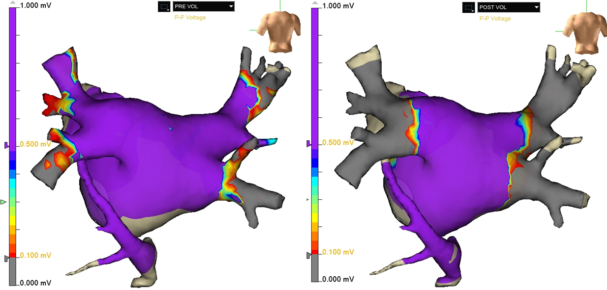
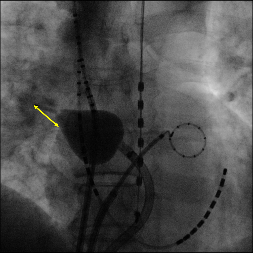
Introduction: Pulmonary vein (PV) isolation (PVI) with a balloon-based visually guided laser ablation (VGLA) is a useful tool for treating atrial fibrillation (AF), however, phrenic nerve injury (PNI) is an important complication. We investigated the predictors of developing PNI during VGLA. Methods and Results: This study included 130 consecutive patients who underwent an initial VGLA of non-valvular paroxysmal AF. During the ablation of the right-sided pulmonary veins, continuous and stable right phrenic nerve pacing was performed, and the compound motor action potentials (CMAPs) were recorded. Twenty patients developed PNI during the PVI. The patients who suffered from PNI had a significantly larger right superior PV (RSPV) ostium area (284.7 ± 47.0 mm2 vs. 233.1 ± 46.4 mm2, P < 0.01) than that of the other patients. Receiver operating characteristic analyses revealed that the area under the curve of the RSPV ostial area was 0.79 (95% confidence interval: 0.69-0.90) with an optimal cut-off point of 238.0 mm2 (sensitivity: 0.58, specificity: 0.95). In the multivariate analyses, large RSPV ostial area (HR 1.02, 95% confidence interval: 1.01-1.03, P < 0.01) and small balloon size (HR 0.72, 95% confidence interval: 0.53-0.98, P = 0.03) were independent risk factors for PNI. PNI remained in 13 patients after the procedure, but 12 of those patients recovered from PNI during the follow-up period. Conclusion: The incidence of PNI during the VGLA was relatively high, but the PNI improved in the majority of cases. During the VGLA, a large RSPV and small balloon size were predictors of PNI.